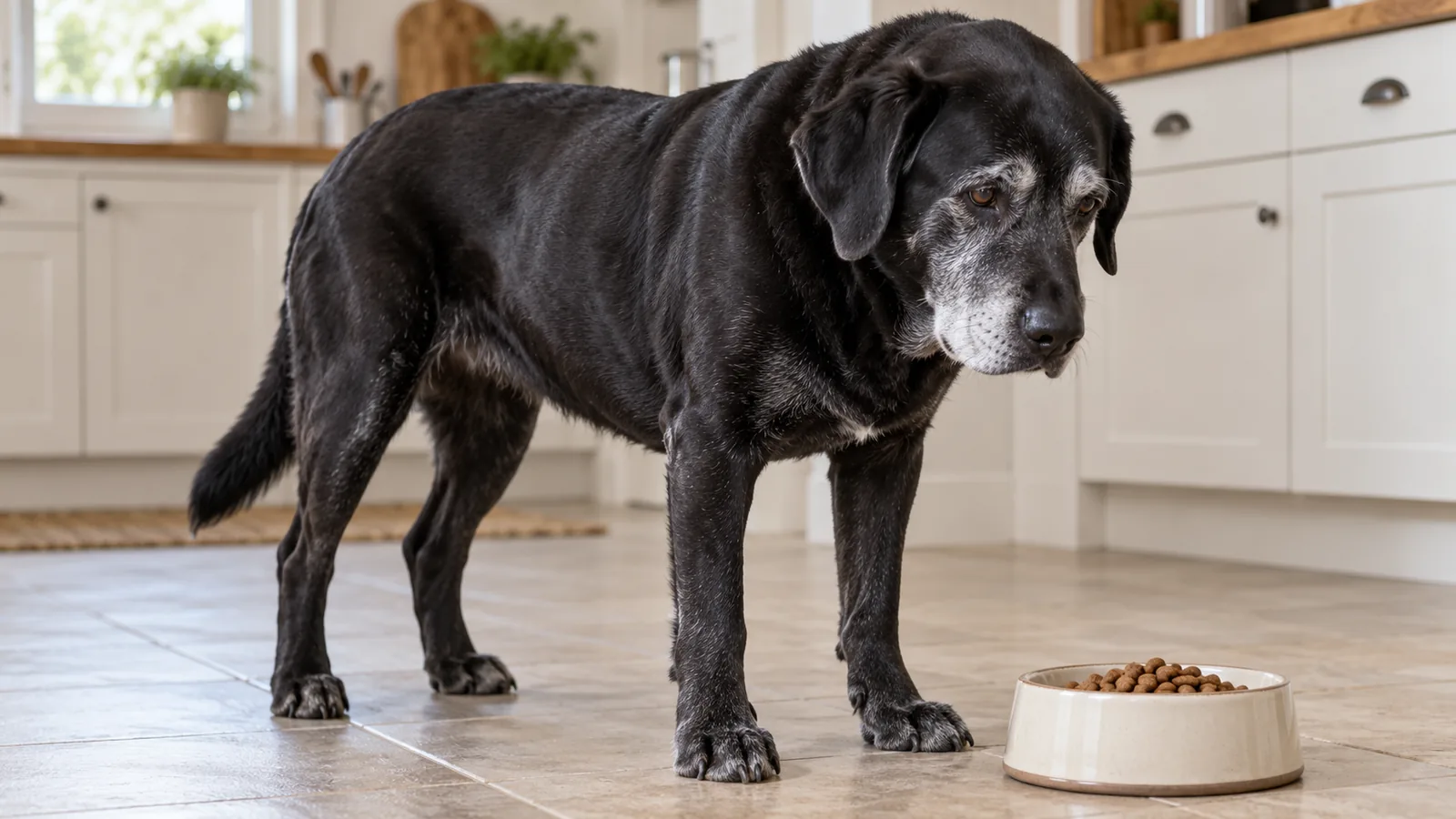
A falling albumin with no hemorrhage, no hepatic failure, and no inflammatory driver to explain it usually means plasma protein is escaping through the intestinal mucosa or across the glomerular filter. Protein-losing enteropathy is usually nonselective, so albumin and globulin fall together, though concurrent inflammatory or infectious disease can keep the latter normal or even raise it. Glomerular loss is selective early, so globulin stays intact while proteinuria climbs on a UPC, which makes that pattern a starting point rather than a verdict. Either syndrome can run quietly, PLE as months of weight loss or intermittent diarrhea and PLN as nothing more than a rising ratio on a wellness panel, and the patients who reach you with ascites, pleural effusion, or peripheral edema are the ones whose oncotic pressure has already fallen far enough to declare itself.
A case like this does not have to leave your practice to get specialist input, and that is the gap VESPECON was built to close. Our board-certified internal medicine specialists can help you localize the loss and build the plan around it: RAAS inhibition and blood pressure control for the glomerular cases, immunosuppression where biopsy supports an immune-mediated enteropathy or glomerulopathy, plus thromboprophylaxis, diet, and treatment of the underlying cause. We stay with the case through ongoing specialty consultations as it evolves, and when a scope or inpatient management is the right next step, we help procure the appointment from our hospital network. If you have a hypoalbuminemic patient who is losing ground, send us the case.
Protein Loss in Dogs and Cats: Four Things to Know First
- Protein-losing enteropathy sheds albumin and globulin together through the intestinal mucosa, while protein-losing nephropathy spares globulin early and presents as persistent renal proteinuria.
- Both syndromes can run quietly until falling oncotic pressure produces ascites, pleural effusion, or peripheral edema.
- Localizing the route of loss comes first, but the specific underlying disease decides whether immunomodulation, fat restriction, chemotherapy, or RAAS blockade is the right lever.
- Loss of antithrombin alongside albumin makes both syndromes prothrombotic, so antithrombotic therapy and close monitoring belong in the plan from the start rather than after an event.
How Do You Separate Intestinal From Glomerular Protein Loss?
The protein profile does most of the sorting. Panhypoproteinemia points at the gut, while hypoalbuminemia with globulins still in range shifts suspicion toward the glomerulus, and urine protein quantification on an inactive sediment separates true glomerular loss from pre-renal and post-renal proteinuria.
Before either syndrome takes the blame, the other causes of a low albumin have to come off the list: hemorrhage, hepatic synthetic failure, third-space loss into a body cavity, exudative skin disease over a large surface, and protein-calorie malnutrition in a patient who hasn't been eating. A UPC sitting modestly above the reference range with active sediment is a different problem from one in the double digits on a quiet sample, and generally only the second drops albumin on its own. The two routes can also coexist, and a patient with both patterns needs a workup for each.
| Feature | Protein-losing enteropathy | Protein-losing nephropathy |
| Route of loss | Intestinal mucosa and lymphatics | Glomerular filtration barrier |
| Protein pattern | Albumin and globulin fall together | Albumin falls, globulin spared early |
| Confirming measurement | Fecal alpha-1 proteinase inhibitor where available, plus serum protein fractions | UPC on repeat samples with inactive sediment |
| Typical presentation | Intermittent diarrhea, weight loss, ascites | Proteinuria on a wellness panel, hypertension, edema |
| Tissue diagnosis | Intestinal biopsy, endoscopic or surgical | Renal biopsy with specialized handling |
| First therapeutic lever | Diet and immunomodulation, or chemotherapy for lymphoma | RAAS blockade and blood pressure control |
What Causes Protein-Losing Enteropathy, and How Is It Confirmed?
Protein-losing enteropathy is a consequence rather than a diagnosis, and the drivers behind protein-losing enteropathy in dogs come down to three. The shared mechanism is mucosal damage or lymphatic obstruction that lets albumin and globulin escape into the lumen, and which one is responsible decides where the plan starts. Yorkshire Terriers, Soft Coated Wheaten Terriers, and Norwegian Lundehunds are overrepresented in this population.
- Chronic inflammatory enteropathy: any of the chronic enteropathies can progress to the point where plasma protein leaves the circulation faster than the liver can replace it.
- Intestinal lymphangiectasia: obstructed intestinal lymphatics that spill protein-rich lymph into the lumen.
- Infiltrative disease: alimentary lymphoma heads this group, and it's the differential that makes a tissue diagnosis matter before immunosuppression goes in.
Clinical signs are often mild enough to be explained away. A dog with soft stool every few weeks and a slow drop in body weight reads as a diet problem until the panel comes back, and in some patients the first hard flag is pitting edema or a fluid wave found at a routine exam. Once panhypoproteinemia appears, PLE diagnosis is largely a process of exclusion, with the differentials above cleared before the workup commits to intestinal biopsy.
Abdominal ultrasound characterizes mucosal detail, wall layering, and the mesenteric lymph nodes, and the radiologists behind our tele-radiology service can read survey films for effusion or an obstructive pattern. Cats are underrepresented in the PLE literature rather than spared, and protein-losing enteropathy in cats usually sits behind small cell lymphoma or chronic enteropathy, with little more than weight loss and intermittent vomiting to announce it.
How Is Advanced PLE Managed?
Management has two jobs: treat the disease driving the loss, and slow the loss itself. For intestinal lymphangiectasia, the highest-yield intervention in slowing intestinal protein loss is a consistently fed ultra-low-fat diet, which often recovers more albumin than another increment of immunosuppression.
Where biopsy supports an immune-mediated or inflammatory enteropathy, immunomodulation takes the lead, usually a glucocorticoid with a steroid-sparing agent added when the response stalls or the side effects become their own problem.
- Cobalamin: supplement it where ileal disease has cost absorption.
- Ionized calcium: measure it rather than inferring from a total value that hypoalbuminemia has already pulled down. Vitamin D malabsorption drives that deficit, and it's worth correcting.
Change one thing at a time, with a scheduled recheck before the next goes in. The albumin trend, not a single value, tells you whether any of it is working. Our clinical advisors in internal medicine can help sequence the diet trial, the immunosuppressive escalation, and the antithrombotic plan for a dog whose protein is still falling.
What Confirms Protein-Losing Nephropathy?
Protein-losing nephropathy is confirmed by a urine protein-to-creatinine ratio that stays elevated on repeat samples with an inactive sediment, in a patient with no inflammation, infection, or urinary tract hemorrhage to explain it. A single ratio is a screen rather than a diagnosis, so document persistence across samples collected days to weeks apart before the proteinuria drives a treatment decision.
Localizing the loss comes before characterizing the lesion. Glomerular disease spans immune-complex glomerulonephritis, amyloidosis, and glomerulosclerosis, and the distinction matters because immunosuppression is justified in the immune-complex group, and confirming that group takes a properly handled renal biopsy. Whether the biopsy is worth pursuing depends on what the result would change, and that question belongs before anesthesia rather than after.
Two things belong in the workup alongside the ratio:
- Blood pressure: hypertension travels with these cases often enough that it belongs in the same visit as the UPC, taken on a settled patient and repeated before anyone commits to a drug for it.
- A search for the driver: tick-borne infection, dirofilariasis, chronic inflammatory disease, and neoplasia can each feed immune-complex deposition, and treating the cause does more for proteinuria than any renal-protective agent on its own.
Cats develop PLN as well, more often alongside amyloidosis or another systemic process than as primary immune-complex disease. Caught while the patient is still eating, still normotensive, and not yet hypoalbuminemic, this is a disease you can slow.
How Is Advanced PLN Managed?
PLN management works on three fronts: cutting the protein crossing the filter, protecting the renal function that remains, and heading off the thromboembolic and hypertensive complications. Blockade of the renin-angiotensin-aldosterone system and blood pressure control carry most of that load, with antithrombotic therapy running alongside. The response you want is a substantial fall in the UPC without a rise in creatinine.
The RAAS inhibitor dose that gets there varies more than the published starting points suggest. Renal nutrition in these patients balances moderating protein enough to ease glomerular load against the muscle loss that chronic protein depletion is already causing, with omega-3 supplementation and sodium moderation built into the same plan.
Long-term PLN care runs on reassessment, with UPC, creatinine, blood pressure, and albumin rechecked within weeks of any medication change rather than at the next annual visit. Several findings along the way are ordinary parts of the course rather than evidence the plan failed:
- Aldosterone escape.
- A creeping creatinine after an ACE inhibitor or angiotensin receptor blocker goes in.
- Hypertension that only partly answers to the first agent.
Where the UPC stays high and albumin keeps dropping through all of that, the question swings back to whether the glomerular lesion itself has ever been characterized.
Why Do These Patients Deteriorate Suddenly?
The deterioration that ends these cases is often thromboembolic rather than gradual. Optimizing outcomes in advanced PLE means treating the prothrombotic state as an active problem from the first visit, not after a pulmonary thromboembolism or aortic thrombus declares itself.
| Complication | What drives it | What it changes in the plan |
| Thromboembolism | Loss of antithrombin and other regulatory proteins, with a relative rise in procoagulant activity | Antithrombotic therapy from the outset, and a low threshold for imaging acute dyspnea or paresis |
| Effusion and edema | Oncotic pressure falling past what the interstitium can compensate for | Cautious diuresis, treatment of the protein loss itself, drainage only when ventilation is compromised |
| Chronic protein depletion | Months of negative nitrogen balance, often with malabsorption on top | Deliberate calorie and protein targets, muscle condition scored at every recheck |
A patient with acute dyspnea, new hindlimb paresis, or sudden collapse needs emergency stabilization at the nearest 24-hour veterinary hospital before anything else, and that call doesn't wait on a consult. For one stable enough to work up, an echocardiogram helps answer whether right heart strain or pulmonary hypertension is part of the picture. The rest is vigilance: a resting respiratory rate recorded at home, a baseline for how the hind end looked last week, and a low threshold for taking a new lameness seriously in a hypoalbuminemic dog or cat.
When Should a Protein-Loss Case Get Specialist Input?
Specialist input helps most before a protein-loss case decompensates. A patient with a localized route of loss but no tissue diagnosis, or one whose albumin keeps falling on the initial plan, is the case to send early rather than after effusion or a thromboembolic event forces the decision.
Three questions separate a specialty-managed case from one that drifts: whether a biopsy will change anything, how to balance competing demands, and how fast to move, because these cases can turn inside a week. Each of those competing demands carries a wrong answer in both directions: immunosuppressing a dog who might have lymphoma, diuresing a patient whose perfusion is already marginal, restricting protein in an animal losing muscle.
What Referring Veterinarians Ask Us About Protein Loss
Can PLE and PLN occur in the same patient?
Concurrent intestinal and glomerular loss is uncommon but real, and it tends to surface when a dog with confirmed chronic enteropathy also carries a UPC that stays elevated on an inactive sample. Quantify the urinary loss, check whether globulin is behaving the way the intestinal picture predicts, and work up each route on its own. Treating only one leaves albumin falling for a reason nobody has addressed.
Why does hypoalbuminemia matter beyond the number on the panel?
Albumin holds water inside the vessels, but it also carries calcium, drugs, hormones, and bilirubin, and it leaves alongside antithrombin. A value of 1.5 g/dL isn't only a prediction of effusion: it's altered drug binding, a total calcium reading falsely low, and a patient primed to clot.
What's a realistic prognosis once a case is advanced?
Outlook in advanced disease is guarded and depends more on the underlying driver than on the lowest albumin recorded. Dogs with intestinal lymphangiectasia or an immune-mediated enteropathy that responds to diet and immunomodulation can hold remission for years, while alimentary lymphoma and amyloidosis carry much shorter horizons. Glomerular cases still non-azotemic when proteinuria is caught do better than those already losing function. Thromboembolism can undo an otherwise decent prognosis, which is why it gets managed before it happens.
Partnering on Complex Protein-Loss Cases
Most of this work happens in general practice, and it goes better with a specialist in the conversation early. VESPECON consultants are available around the clock, so if you have a hypoalbuminemic dog or cat and a route of loss you want a second read on, reach out about the case while the patient is still in front of you.